HIP dysplasia causes premature arthritis and HIP pain in young adults, but for many patients, the natural HIP can be preserved with pelviacetabular surgery by a pelvic fracture specialist. Correct the socket before arthritis is irreversible. Delay or avoid total HIP replacement by decades.
Our specialist will call you within 2 hours
HIP dysplasia is a condition in which the acetabulum (the socket of the HIP joint) is too shallow or incorrectly oriented — failing to provide adequate coverage of the femoral head (ball). This instability causes abnormal load distribution across the joint, accelerated cartilage wear and, if untreated, progressive HIP arthritis in young adults. Many patients with HIP dysplasia are not diagnosed until their 20s or 30s, when HIP pain, clicking and functional limitation prompt investigation.
Pelviacetabular surgery — most commonly the Periacetabular Osteotomy (PAO) — is a HIP preservation procedure designed to correct the underlying anatomical problem before arthritis becomes irreversible. The acetabular bone is carefully cut, repositioned to provide optimal coverage of the femoral head, and fixed in the corrected position with screws. The goal is to distribute load normally across the joint, eliminate instability and stop the progression of arthritis — preserving the patient’s own HIP for decades and, in many cases, avoiding or significantly delaying total HIP replacement.
HIP dysplasia is frequently undiagnosed until early adulthood — these are the symptoms and findings that should prompt specialist assessment.
Deep groin pain in a patient aged 15–40 — especially with activity, prolonged standing or sitting — is the classic presentation of HIP dysplasia. Young adults with groin pain are often investigated for muscle strain or sports injuries before the HIP joint is imaged. A standing AP pelvis X-ray is the essential first investigation.
A clicking or clunking sensation in the HIP with certain movements — particularly HIP flexion and internal rotation. In a dysplastic HIP, this represents the unstable femoral head moving abnormally within the shallow socket. Often mistaken for a soft tissue problem — it is a structural joint problem.
Pain during or after running, sports, prolonged walking or cycling — that was not present a few years earlier or has progressively worsened. Activity-related HIP pain in a young adult that does not resolve with physiotherapy is a red flag for underlying structural pathology requiring imaging.
A limp present since early childhood or noticed progressively through adolescence — often attributed to muscle weakness or growing pains. Many adult patients with HIP dysplasia have a history of a "clicky HIP" as an infant or a limp that was never fully investigated. This history should always prompt HIP X-ray in adult assessment.
Standing AP pelvis X-ray showing a shallow acetabulum — reduced lateral centre-edge angle (under 20 degrees), acetabular inclination above 10 degrees or visible undercoverage of the femoral head. These radiological findings confirm dysplasia and quantify its severity — providing the basis for surgical planning.
MRI showing a HIP labral tear in a young patient — the labrum is the cartilage seal around the rim of the acetabular socket. In a dysplastic HIP, the labrum is subjected to abnormal load and tears early. A labral tear in a young adult should always prompt a standing AP pelvis X-ray to exclude underlying dysplasia as the cause.
A first-degree relative with developmental dysplasia of the HIP (DDH) or HIP replacement at a young age significantly increases the risk. Patients with a family history of DDH who develop HIP pain should be assessed with standing X-rays earlier rather than later — the window for HIP preservation surgery closes as arthritis progresses.
Joint space narrowing or early arthritic change on HIP X-ray in a patient under 40 — findings that are unexpected for age and should prompt investigation into an underlying structural cause. HIP dysplasia is the most common structural cause of early-onset HIP arthritis. Assessment for correctable dysplasia should occur before cartilage loss is too advanced for preservation surgery.
Pelviacetabular surgery requires specific expertise — not every orthopaedic centre offers PAO or has the case volume to deliver consistent outcomes.

HIP Preservation & Pelviacetabular Surgery, Trayam Hospital
Dr. Parth Patel is a fellowsHIP-trained HIP preservation surgeon with specific expertise in periacetabular osteotomy, acetabular reconstruction and the management of HIP dysplasia in adolescents and young adults. The central philosophy is straightforward: for a young patient with a dysplastic HIP and preserved cartilage, correcting the socket before arthritis becomes irreversible gives the best long-term outcome. Total HIP replacement in a 30-year-old is a last resort — not a first answer — and everything that can be done to delay or avoid it should be attempted first.
The right procedure depends on patient age, degree of dysplasia, cartilage status and whether arthritis has already developed.
A step-by-step guide to your recovery after Pelvi Acetabular Surgery at Trayam Hospital.
Surgery completed. Strict bed rest on day 1. Physiotherapy begins day 2 — gentle range-of-motion exercises and deep breathing. Toe-touch weight-bearing only initially.
Hospital discharge. Walking with crutches — protected weight-bearing for 6–8 weeks while osteotomy heals. Pain managed with oral medication. X-ray confirms osteotomy position before discharge.
Follow-up X-ray assessing osteotomy healing. Physiotherapy progressing — strengthening exercises, range of motion improving. Most patients managing light daily activities from home.
X-ray confirming osteotomy healing — progression to full weight-bearing. Crutches weaned. Physiotherapy entering strengthening and gait rehabilitation phase.
Walking normally without aids. Return to desk work and light daily activities. Significant improvement in groin pain and HIP function compared to pre-surgery. Physiotherapy continuing with strength and functional goals.
Return to recreational sport and full activity for most patients. Final outpatient review with X-ray confirming complete osteotomy union and implant position. Annual review recommended to monitor HIP joint health long-term.
HIP dysplasia is one of the most consistently under-diagnosed conditions in orthopaedics. Young adults — particularly women — with groin pain, HIP clicking and activity limitation are frequently investigated for muscle injuries, sports hernias and soft tissue problems for months or years before a standing pelvis X-ray is obtained. By the time the diagnosis is made, some patients have progressed to early arthritis that narrows or closes the window for HIP preservation surgery. Early diagnosis and early referral to a HIP preservation specialist is the single most important factor in determining whether PAO is possible.
Every patient referred for HIP pain assessment receives a standing AP pelvis X-ray with standardised measurements. If dysplasia is confirmed, CT is arranged for 3D planning. The diagnosis drives the decision — not a default recommendation.
If PAO is possible given your age and cartilage status, we will offer it. Total HIP replacement in a young patient is a last resort — not a first answer. We will tell you honestly whether the window for preservation is open or closed.
PAO and pelviacetabular reconstruction are covered under most major health insurance policies. We manage the pre-authorisation process in full, ensuring that the procedure and its indication are correctly documented for your insurer.
Young adult with HIP pain? Shallow socket on X-ray? HIP dysplasia diagnosed? Come in early — the window for preservation closes with time.
Confidential · No Obligation · Reply in 2 Hours
HIP dysplasia is a condition where the socket does not fully cover the femoral head, leading to instability and early arthritis. In many cases, surgical correction by a pelvic fracture specialist in Ahmedabad helps restore joint stability and prevent long-term damage.
PAO is a surgical procedure in which the acetabulum is cut free from the pelvis at four points, repositioned to provide optimal femoral head coverage and fixed in the corrected position with screws. It is the gold standard HIP preservation procedure for symptomatic dysplasia in patients under 40 with preserved cartilage.
Ideal candidates are under 40, have symptomatic HIP dysplasia confirmed on standing X-ray, and have preserved or near-preserved cartilage on MRI arthrogram. Patients with advanced arthritis (Tönnis grade 3 or above) are generally not candidates for PAO and are better served by total HIP replacement.
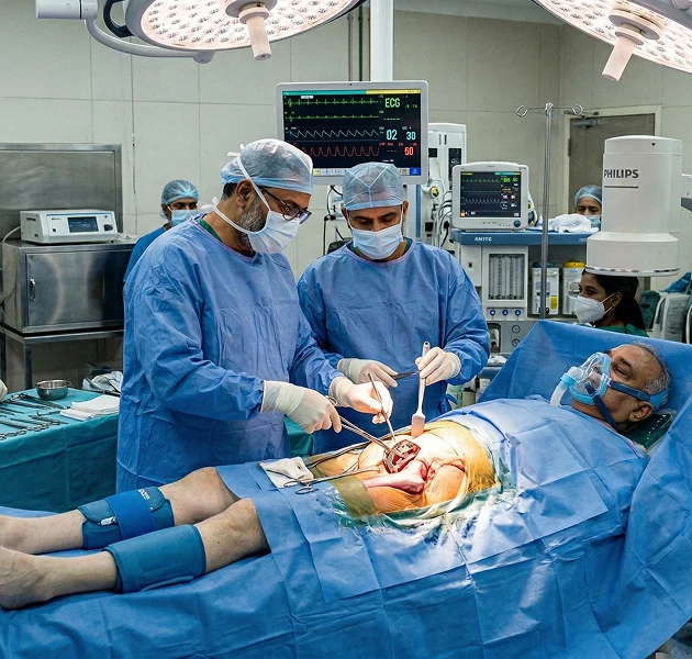
The procedure takes approximately 2–3 hours under general anaesthesia. Hospital stay is typically 5–7 days.
Protected weight-bearing on crutches for 6–8 weeks while the osteotomy heals. Walking without aids by 8–10 weeks. Return to light activity at 3–4 months. Return to sport at 6–12 months. Full recovery assessment at 12 months.
In correctly selected patients, PAO significantly delays or avoids total HIP replacement. Long-term outcome studies show over 70% of PAO patients retain their natural HIP at 20 years. The earlier the procedure is performed — before significant cartilage loss — the better the long-term outcome.
Physiotherapy can improve HIP muscle strength and reduce symptoms but does not correct the underlying structural problem. Progressive cartilage damage continues even when symptoms are partially managed with physiotherapy. In patients with significant dysplasia and symptoms, surgical correction is the only way to address the cause.
Yes — periacetabular osteotomy and pelviacetabular surgery are covered under most major Indian health insurance policies. Our insurance desk manages the pre-authorisation process and cashless hospitalisation in full.
The earlier we assess, the more options are available. Bring your X-rays or MRI if you have them — or we arrange a standing pelvis X-ray to start. Free consultation, no obligation.